The TMIG Standard Procedure of 2-D PAGE in Proteomics
Tosifusa Toda
Proteomics Collaboration Center
Tokyo Metropolitan Institute of Gerontology
Last update : Nov 1, 2007
Please cite the following our papers in your paper if you use our
optimized procedures for proteomic analysis.
And we also recommend you to refer to this URL (http://proteome.tmig.or.jp/2D/2DE_method.html),
because the procedures shown in this Web site include slight
modifications and supplements.
Related papaers on applications of our 2-D gel-based proteome
analysis
Apparatus
- First dimension : immobilized pH-gradient isoelectric
focusing
CoolPhoreStar IPG-IEF (Anatech) 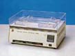
Protean IEF Cell (Bio-Rad) 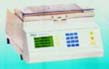
- Second-dimension : SDS-PAGE
CoolPhoreStar SDS-PAGE Tetra-200 (Anatech) 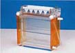
Protean II XL (Bio-Rad) 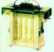
- The source of electric power
PowerPhoreStar Pro3800 (Anatech, Japan) 
Powerpack 3000 (Bio-Rad) 
Reagents and Consumables
Tris (TRIZMA BASE),
- Sigma: T-1503
Urea,
- Nakarai Tesque: Code No. 35940-81
Thiourea,
- Sigma: T-7875
CHAPS,
- Sigma: C-5849
Tricine,
- Sigma: T-0377
Sulfobetain 10 (SB3-10),
- Amresco: J548-10G
Orange G,
- Sigma: O-1625
Electrofocusing sample application pieces,
- Amersham Pharmacia Biotech: Code No. 80-1129-46
- or Anatech: Code No. 3600-13
Electrofocusing electrode wicks,
- Amersham Pharmacia Biotech: Code No. 18-1004-40
- or Anatech: Code No. 3600-12)
Silver stainning kit,
- Wako Pure Chem.: Code No. 299-13841
Silcon oil 5c/s,
- Shin-Etsu
Chem.: Code No. KF-96-L-5CS
pI Marker protein kit,
- Daiichi Chem.: Code No. 220760
Molecular weight marker protein kit,
- Daiichi Chem.: Code No. 181122 or 181066
Gel strip for the first-dimensional
IPG-IEF (Immobilized pH gradient isoelectric focusing):
Rehydrate Immobiline DryStrip (Amersham Pharmacia Biotech) or IPG
ReadyStrip (Bio-Rad) using a rehydration solution, such as shown below:
urea                  14.4 g
thiourea               6.1 g
dithiothreitol         0.08 g
Pharmalyte 3-10        0.4 ml
0.1 M acetic acid      1.0 ml
0.1%(w/v) Orange G     1.0 ml
20%(v/v) Triton X-100  4.0 ml
pure water     up to  40.0 ml

The volume is for 8 strips of 18-cm or 17-cm IPG gel (5 ml per strip).
Gel plates for the second-dimensional SDS-PAGE:
35 %(w/v) acrylamide         106  ml
 2 %(w/v) BIS-acrylamide      58  ml
1.5 M     Tris-HCl, pH 8.8   124  ml
60%(v/v)  glycerol            90  ml
pure water    up to          500  ml
--- degas in vacuo before mixing reagents shown below---
10 %(w/v) SDS                 5.0 ml
10 %(w/v) ammonium persulfate 1.0 ml
TEMED                         0.3 ml

The volume is for 8 plates of 7.5%T, 3%C polyacrylamide gel (190 x 180 x 1 mm).
Preparation of protein extraction reagents:
   For cytosolic/hydrophilic proteins       For membrane/particulate-bound proteins
   Urea                   0.51  g           Urea                   0.30  g
   10%(w/v) SDS           0.02 ml           thiourea               0.15  g
   10%(v/v) Triton X-100  0.20 ml           20%(w/v) CHAPS         0.10 ml
   DTT                    0.01  g           20%(w/v) SB3-10        0.10 ml
   Pharmalyte 3-10        0.02 ml           DTT                    0.01  g
   Milli-Q water    up to 1.00 ml           Pharmalyte 3-10        0.02 ml
                                            Milli-Q water    up to 1.00 ml

   [Supplements for preventing protein degradations completely (Optional)]
    Sigma Proteinase inhibitor Cocktail   10 ul/ml
    Sigma Phosphatase Inhibitor I          5 ul/ml
    Sigma Phosphatase Inhibitor II         5 ul/ml
Preocedure of protein extraction from
tissue or packed cell pellet:
- Suspend tissue fragments or cells on 3 volumes of Protein
Extraction Reagent A or B.
- Disrupt cells by ultrasonication for 1 sec x 20 times.
- Remove the supernatant by centrifugation at 15,000 rpm for 20
min at room temperature.
- Store small aliquots of the supernatant below -70C until use
for electrophoresis. (Incubate for 3 min at 45C prior to application)
Protein extraction from cell cultures
(Alternative ):
- Rinse the culture dish with 5 ml of PBS- for 1 min, repeat 3
times.
- Scrape off cells from the culture dish on ice by rubbing with a
plastic scraper.
- Transfer the cell suspension to a 1.5-ml microfuge tube of
which tare was weighed in advance. (Give W [mg]= the weight of the cell
suspension)
- Add W x 0.85 mg of urea, W x 0.05 micro-liter
2-mercaptoethanol, W x 0.1 micro-liter of 1.5%(w/v) SDS, 8.5%(v/v)
Triton X-100.
- Disrupt cells by ultrasonication for 1 sec x 20 times.
- Remove the supernatant by centrifugation at 15,000 rpm for 20
min at room temperature.
- Store small aliquots of the supernatant below -70C until use
for electrophoresis. (Incubate for 3 min at 45C prior to application)
Procedure of the first-dimensional IPG-IEF
(Immobilized pH-gradient isoelectric focusing):
- Pour a small volume of silicon oil into the electrophoresis
chamber, mount Immobiline DryStrip supporting plate in the chamber
giving care to remove air bubbles beneath the plate.
- Add 1 micro-liter of pI marker protein solution to an aliquot
(10-20 micro-liter) of cell extract if neccessary. (In the case of
"DAIICHI" 2D pI Marker for silver staining, the content of a vial is
dissolved in 0.5 ml of pure water. The reconstituted solution is
storable at -20C ).
- Stand re-swelled Immobiline Dry Strip on its long edge on
filterpaper briefly to remove excess Gel Swelling Solution, then lay on
the filterpaper gel-side-up.
- Put a piece of Sample Application Filter on the gel ca. 1-cm
inside from the cathodic* end. Apply the cell extract (10-20
micro-liter) to the Sample Application Filter.
(* This is for pH 4-7 and 3-10 Drystrips. Anodic application is better
for pH 6-11 Drystrip)
- Mount the strip in the electrophoresis chamber which is
pre-cooled to 20C.
- Put electrode pads wetted with distilled water on both ends of
the strip to recieve sure contact of platinum electrodes.
- Set the platimum electrode, and cover the gel strip and
electrode pads with silicon oil (5 c/s) to isolate electrophoresis
media from the atmosphere.
- Apply electric power in the stepwise elevation of volutage as
follows:
For pH 4-7 and 3-10 Drystrips		For pH 6-11 DryStrip  For pH 5.0-6.0 DryStrip

	(18-cm long)			(18-cm long)			(18-cm long)

 Step	Voltage	Time (h)		Voltage	Time (h)		Voltage	Time (h)

  1	 500 V	 2 h			 350 V	8 h			 500 V	 1.0 h
  2	 700 V	 1 h			 500 V	1 h			 700 V	 0.5 h
  3	1000 V	 1 h			1000 V	1 h			1000 V	 0.5 h
  4	1500 V	 1 h			1500 V	1 h			1500 V	 0.5 h
  5	2000 V	 1 h			2000 V	1 h			2000 V	 0.5 h
  6	2500 V	 1 h			2500 V	1 h			2500 V	 0.5 h
  7	3000 V	 1 h			3000 V	1 h			3000 V	 0.5 h
  8	3500 V	10 h			3500 V	3 h			3500 V	13.0 h
Keep the electric power supply at 500 V after the step 8.
Treatment of the gel strip for the
second-dimensional SDS-PAGE:
1. Primary treatment for disulfide reduction.
Incubate the gel strip for 30 min at room temperature in the
SDS-treatment solution prepared as follows.
2. Secondary treatment for alkylation.
Incubate the gel strip for 20 min at room temperature
in one of the following alkylation reagents.
Carbamoylmethylation (CM) Reagent for 8 strips:

0.5 M  Tris-HCl, pH 6.8    2.0 ml
10%(w/v) SDS stock         8.0 ml
0.1%(w/v) BPB              1.0 ml
60%(v/v) glycerol         20.0 ml
Milli-Q water              9.0 ml
iodoacetamide              1.8 g             ICH2CONH2
                       --> Protein-S-CH2-CO-NH2
or
Pyridylethylation (PE) Reagent for 8 strips:
0.5 M Tris-HCl, pH 6.8 2.0 ml
10%(w/v) SDS stock 8.0 ml
0.1%(w/v) BPB 1.0 ml
60%(v/v) glycerol 20.0 ml
Milli-Q water 9.0 ml
4-vinylpyridine 1.0 ml CH2=CH-Pyridyl
--> Protein-S-CH2-CH2-Pyridyl
Procedure of the Second-dimensional SDS-PAGE
- Pour an appropriate volume of the anodic electrode solution
into the vertical electrophoresis chamber.
- Incubate the gel strip in SDS-treatment solution for 5 min at
room temperature again.
- Fill the top of the gel slab with the cathode solution, place
the gel strip that is trimmed to fit the gel size, and gently push the
strip down using a shark tooth-shaped Strip Fixer Comb to achieve the
firm contact to the gel slab.
- Mount the gel plate on the electrophoresis chamber unit.
- Fill the cathodic chamber with the cathodic electrode buffer.
- Perform electresis in 20-40 mA/slab until the BPB dye comes
near the gel bottom.
- Proceed to the subsequent protein staining or an
electro-transfer blotting.
Electrode Buffer Solutions for SDS-PAGE:
Preparation of 10x stock of anodic electrode
buffer (2 M Tris-HCl, pH 8.8):
Tris      242.2 g
in 700 ml of Milli-Q SP water.

Adjust pH to 8.8 with 6N HCl
Add pure water upto 1,000 ml
Preparation of 5x stock of cathodic electrode
buffer (Tris-Tricine Buffer ):
Pro-Q Diamond Staining for phosphoprotein
detection
- Immerse the gel in 50%(v/v) methanol, 10%(v/v) acetic acid and
incubate for at least 30 min at room temperature.
- Repeat the fixaton step once more to remove SDS from the gel
completely. Gel can be left in the fixing solution overnight.
- Rinse the gel in Milli-Q water for 10 min.
- Repeat the rinsing once more to remove methanol and acetic acid
completely. Residual methanol or acetic acid will interfere with Pro-Q
Diamind staining.
- Immerse the gel in Pro-Q Diamond phosphoprotein gel stain with
gentle agitation for 75-120 min in the dark.
- Rinse the gel in the Destain Solution for 1 h in the dark.
- Repeat the rinsing once more.
For preparation of Destain Solution:
- Add 50 ml of 1 M sodium acetate, pH 4.0, and 200 ml of
acetonitrile to 750 ml of
Milli-Q water.
SYPRO Ruby Staining
- Incubate the gel in 50%(v/v) methanol, 10%(v/v) acetic acid for
more than 30 min. Gel can be left in the fixing solution overnight.
- Incubate the gel in 10%(v/v) methanol, 7%(v/v) acetic acid for
30 min.
- Immerse the gel in the dark in SYPRO Rubu gel stain with gentle
agitation for 90-120 min.
- Rise the gel in 10%(v/v) methanol, 7%(v/v) acetic acid for 30
min.
Coomassiee Blue Staining (optinal)
- Incubate the gel in 50%(v/v) methanol, 10%(v/v) acetic acid for
more than 30 min.
- Incubate the gel in working reagent of Quick-CBB Protein Stain
Reagent Kit (299-50101, Wako Pure Chemicals) for 30 min - 1 h.
- Rise the gel in 7%(v/v) acetic acid.
Silver staining (optinal)
In our institute, Silver Stain Kit "DAIICHI" (708104-002, Daiichi Pure
Chemicals) is generally used.
(Note) The step for protein fixation should be skipped if the protein
should be resolubilized and transffered to PVDF membrane after silver
staining.
In-Gel Protein Digestion for Peptide Mass
Fingerprinting
- Pick a small gel piece up from a protein spot, and put it in a
96-well PCR plate (ABGene Thermo-Fast 96, Skirted).
- Immerse the gel piece in 0.1 ml of Reducing Reagent for 30 min.
- Reducing Reagent :1.5 mg/ml dithiothreitol in 100 mM ammonium
bicarbonate
- Immerse the gel piece in 0.1 ml of Alkylating Reagent for 30
min.
- Alkylating Reagent :10 mg/ml iodoacetamide in 100 mM ammonium
bicarbonate
- Immerse the gel piece in 0.1 ml of 50% methanol, 50mM ammonium
bicarbonate for 15 min x 2.
- Immerse the gel piece in 0.1 ml of 50% acetonitrile, 50mM
ammonium bicarbonate for 10 min x 3.
- Immerse the gel piece in 0.1 ml of 100% acetonitrile for 5 min.
- Remove the liquid completely and dry up the gel.
- Reswell the gel piece in 20 ul of the Digestion Reagent.
- Digestion Reagent: 5 ug/ml sequencing grade modified trypsin
(Promega V511A or Sigma T-6567),
- in 30%(v/v) acetonitrile, 50 mM ammonium bicarbonate.
- Seal up the PCR plate with an adhesive seal (SUMILON Plate Seal
MS-30010, Sumitomo Bakelight Co. Ltd.) to prevent evaporation.
- Incubate the gel piece in the digestion reagent overnight at
30C.
* Extra extraction procedure is not necessarily required for
MALDI-TOF/MS.
Sufficient extraction of digested peptides into the digestion reagent
has been finished during the overnight incubation in our digestion
reagent. The digestion reagent after incubation is applicable to a
MALDI target plate by direct mixing with an equivalent volume of matrix
solution.
* Step 2 and 3 can be skipped if the sample protein has already been
alkylated before 2-DE.
Re-solubilization of Stained Proteins in the
Gel and Electrotransfer blotting to PVDF membrane (An alternative
procedure to the
in-gel digestion)*
- Rinse the gel in pure water, for 15 min x 3 times.
- Incubate in the re-solubilization reagent (0.3%(w/v) Tris,
0.7%(w/v) glycine, 0.2%(w/v) SDS) for 15 min.
- Overlay with Sequi-Blot PVDF membrane (Bio-Rad), and mount in a
blotting chamber with the electrotransfer buffer (0.3%(w/v) Tris,
1.5%(w/v) glycine, 0.1%(w/v) SDS)
- Apply 4 V/cm in the electrotransfer buffer (0.3%(w/v) Tris,
1.5%(w/v) glycine, 0.1%(w/v) SDS) overnight.
*Please cite our previous paper "Proteomic
analysis of Epstein-Barr virus-transformed human B-lymphoblastoid cell
lines before and after immortalization" Toda T, Sugimoto M, Omori A,
Matsuzaki T, Furuichi Y, Kimura N., Electrophoresis. 2000
May;21(9):1814-22, if you use the procedure for electrotransfer
blotting after staining of protein in your research.
On-Membrane Protein Digestion
- Punch out a small area of protein spot from a stained
Sequi-Blot PVDF membrane (Bio-Rad).
- Put it in a microfuge tube siliconized with SIGMACOTETM.
- Destain the membrane with 60%(v/v) methanol, 10%(v/v) acetic
acid (15 min x 3).
- Bufferize the membrane in 2.5 mM Tris-HCl, pH 7.7 (3 min x 3).
- Treat the membrane with 4.5%(w/v) Polyvynyl pirolidone 25, 2.5
mM Tris-HCl, pH 7.7 for 30 min.
- Wash the membrane with pure water (3 min x 3).
- Wash the membrane with 5%(v/v) acetonitrile (1 min x 3).
- Dip the membrane in 30 micro-l of 8%(v/v) acetonitrile, 2.5 mM
Tris-HCl, pH 7.7.
- Incubate the membrane in 20 micro-l of 0.15 U/ml Endoproteinase
Lys-C (Boehringer Cat#13832451) diluted in 8%(v/v) acetonitrile, 2.5 mM
Tris-HCl, pH 7.7 overnight at 30C.
- Add 20 micro-l of acetonitrile, and incubate for more 1 h at
room temperature.
- Remove the peptide solution to another tube, and desalt using a
ZipTip for MS analysis or microsequencing (optional).
*Please cite our previous paper "Proteomic
analysis of Epstein-Barr virus-transformed human B-lymphoblastoid cell
lines before and after immortalization" Toda T, Sugimoto M, Omori A,
Matsuzaki T, Furuichi Y, Kimura N., Electrophoresis. 2000
May;21(9):1814-22, if you use the procedure for the "on-membrane
digestion" in your research.
Desalting with ZipTip (optional)
- Evoporate the solvent using SpeedVac or under a flow of
nitrogen gas.
- Dissolve peptides in 10 micro-l of 0.1% TFA.
- Adsorb peptides on ZipTip C18 resin (Millipore), that has been
previously prewetted with 50% acetonitrile and equilibrated with 0.1%
TFA.
- Wash the resin with 10 micro-l of 0.1% TFA by 5 strokes of
pumping.
- Elute peptides into 5 micro-l of 50% acetonitrile, 0.1% TFA.
- Mix 1 micro-l of peptide solution with 1 micro-l of matrix
solution, and apply to a target of MALDI-TOF/MS.
Matrix solution for MALDI-TOF/MS
Alpha-cyano-4-hydroxycinnamic acid (Sigma C-2020) 10 mg
Acetonitrile (HPLC grade) 0.5 ml
Methanol (HPLC grade) 0.4 ml
TFA (Sigma T-6508) 1 µl
Milli-Q water 0.1 ml
All rights are reserved by T. Toda
(2000-2007)
Most part of this protocol is based on our standardized procedure
published in Jpn. J. Electroph. 1997, 41(1), 13-19. However, the
current version has been slightly modified for optimization in
proteomics after publication. Please cite this URL
(http://proteome.tmig.or.jp/2D/2DE_method.html) when you refer to our
prodedure in your publication.